Endospore Staining – A Comprehensive Approach
Detailed information on Endospore staining, including its types, Procedure, significance and limitations and FAQs.
What is Mean by Endospore Staining
Endospore staining is a laboratory technique that uses dyes to distinguish endospores from vegetative cells in bacterial samples. Endospores are highly resistant structures produced by some bacteria as a survival mechanism under harsh environmental conditions. These spores are resistant to heat, desiccation, radiation, and chemical disinfectants, making them difficult to eradicate. Endospore staining is an essential laboratory technique in microbiology that allows microbiologist to identify. bacterial species capable of producing endospores.
What is an endospore
An endospore is a dormant structure produced by certain types of bacteria to allow them to survive in adverse conditions.
Types of Endospore Staining
There are several types of this techniques, including the Schaeffer-Fulton staining method, Dorner’s method, and Moeller’s method.
Schaeffer-Fulton Staining
The Schaeffer-Fulton staining method is the most commonly used technique for endospore staining. It involves heating the bacterial sample with malachite green dye, which penetrates the endospores’ tough outer layer. The excess dye is washed off, and the sample is counterstained with safranin or fuchsine, which stains the vegetative cells.
Dorner’s Method
Dorner’s method is a modification of the Schaeffer-Fulton staining method. It involves the addition of 1% tannic acid to the malachite green dye, which increases the dye’s affinity for endospores. The excess dye is washed off, and the sample is counterstained with safranin or fuchsine.
Moeller’s Method
Moeller’s method involves heating the bacterial sample with a mixture of malachite green and basic fuchsin dyes. The excess dye is washed off, and the sample is counterstained with methylene blue or neutral red.
Materials Required for Endospore Staining
Before starting the endospore staining procedure, the following materials are required:
- Bacterial culture containing endospores
- Glass slides
- Inoculation loop
- Bunsen burner or alcohol lamp
- Heat source (incubator or water bath)
- Crystal violet stain
- Gram’s iodine solution
- Ethanol or acetone decolorizing agent
- Safranin counterstain
- Microscope
Endospore Staining Procedure
The endospore staining procedure involves several steps, including the preparation of the bacterial smear, heat fixation, staining, decolorization, and counterstaining.
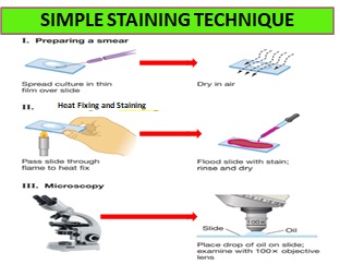
Preparation of Bacterial Smear
The first step is to prepare a bacterial smear by spreading a small amount of the bacterial sample onto a glass slide. The smear is air-dried and then heat-fixed by passing the slide through a flame.
Heat Fixation
Heat fixation is the second step and it involves passing the slide through a flame several times to kill the bacteria, fix them to the slide, and denature their proteins, making them more permeable to the dye.
Staining
Staining is the third step and it involves The bacterial smear is then flooded with malachite green dye and heated over a Bunsen burner for several minutes to allow the dye to penetrate the endospores’ tough outer layer. The excess dye is washed off with water.
Decolorization
The fourth step in the endospore staining procedure is decolorization and Decolorisation is a process of washing the slide with a decolorizing agent, such as water or alcohol, to remove the excess dye from the vegetative cells.
Counterstaining
The final step in the endospore staining procedure is counterstaining. it is the process of staining in which slide is counterstained with a contrasting dye, such as safranin or fuchsine, to stain the vegetative cells, which appear red or pink, while the endospores retain their green color.
Interpretation of Results
Under the microscope, bacterial endospores will appear as green, oval-shaped structures within or near the vegetative cells. The vegetative cells will appear pink or red due to the counterstain. If no endospores are present, all cells will appear pink or red.
Significance of Endospore Staining
It is an important laboratory technique in microbiology because it allows microbiologist to identify bacterial species that are capable of producing endospores. These spores are highly resistant to environmental stressors, making them difficult to eradicate, and can survive for long periods, making them a potential source of contamination. Endospore staining can help identify the presence of endospores in food, water, and medical equipment, and aid in the diagnosis of bacterial infections.
Limitations of Endospore Staining
Endospore staining has some limitations, including:
- Some bacterial endospores may not stain well, resulting in false-negative results.
- Some bacterial endospores may appear as spurious structures, leading to false-positive results.
- It cannot differentiate between different species of endospore-forming bacteria.
- This Technique cannot determine the viability of the endospores.
- The Technique can only detect endospores in bacterial samples, and not in other microorganisms such as fungi, viruses, or protozoa
Safety Considerations while Endospore staining
This staining technique involves the use of flammable liquids, high temperatures, and potentially pathogenic bacteria. Therefore, the following safety precautions should be Taken:
- Wear appropriate personal protective equipment, such as gloves, lab coat, and safety glasses.
- Work in a well-ventilated area.
- Use caution when handling flammable liquids.
- Use caution when working with high-temperature sources, such as Bunsen burners.
- Handle bacterial cultures with care and dispose of them properly.
Troubleshooting
Some common issues while performing the endospore staining and their possible causes include:
- Poor staining: inadequate heat fixation, insufficient staining time, expired stain solutions, or improper decolorization.
- False-positive results: spurious structures mistaken for endospores, or non-endospore-forming bacteria stained as endospores.
- False-negative results: endospores that do not stain well or are not present in the sample, or poor microscope technique.
Frequently Ask Questions (FAQs) :
What is the difference between endospores and vegetative cells?
Endospores are highly resistant structures produced by some bacterial species, which enable them to survive harsh environmental conditions. Vegetative cells are the actively growing and dividing form of bacteria.
What is the purpose of heat fixation? Heat fixation is used to kill the bacteria, adhere them to the slide, and enhance the uptake of the stain.
What is the significance of endospore staining in microbiology?
It is significant in microbiology because it allows microbiologist to identify bacterial species capable of producing endospores, which can be a source of contamination and infection.
What are the types of endospore staining methods?
There are several types of endospore staining methods, including the Schaeffer-Fulton staining method, Dorner’s method, and Moeller’s method.
Why is endospore staining important?
This technique is important for identifying bacterial endospores, which are highly resistant to many disinfectants and antibiotics and can pose a significant challenge for healthcare providers.
Can endospore staining detect endospores in other microorganisms besides bacteria?
No, endospore staining can only detect endospores in bacterial samples, and not in other microorganisms such as fungi, viruses, or protozoa.
What is the purpose of decolorization?
Decolorization is used to remove the stain from the vegetative cells while leaving the endospores stained
What are the limitations of endospore staining?
The limitations of this technique include the inability to detect endospores in non-bacterial microorganisms and the potential for false-negative results in bacterial species that do not produce endospores under certain conditions.