Simple Staining – A simplified Technique for Identification of Microbes

Detailed information on simple staining, including the definition, techniques, procedures, and applications, Limitations and FAQs.
Simple Staining – A Technique for Identification of Microbes
What is Simple Staining?
Simple staining is a staining technique in which a single dye or stain is used to color cells or tissues. The stain used is usually a basic dye, meaning that it is positively charged and will bind to negatively charged cellular components such as nucleic acids and acidic proteins. This technique is used to enhance the contrast of cells or tissues, making them more visible under the microscope.
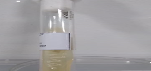
Image I : Simple Staining Technique

Types of Stains used in Simple Staining
There are various types of stains that can be used in this type of staining, Includes,
Crystal Violet
This stain is a deep purple color and is commonly used in Gram staining. It is a basic dye that binds to negatively charged components of bacterial cells, such as peptidoglycan.
Methylene Blue
This stain is commonly used in biology laboratories and is a basic dye that stains nuclei and other acidic cellular components blue.
Safranin
This stain is used in Gram staining and endospore staining. It is a basic dye that stains the cell wall and other structures pink or red.
Basic Fuchsin
This stain is used in acid-fast staining and is a basic dye that stains mycobacteria bright red.
Procedure of Simple Staining
The procedure of this staining method is relatively simple and involves the following steps:
- Prepare a bacterial smear by spreading a thin layer of the bacterial culture on a slide and allowing it to air dry.
- Heat-fix the smear by passing it through the flame of a Bunsen burner or alcohol lamp.
- Flood the slide with the chosen stain, making sure to cover the entire smear.
- Rinse the slide with distilled water to remove excess stain.
- Blot the slide with filter paper to remove excess water.
- Observe the slide under the microscope.
Applications of Simple Staining
This staining method has various applications in microbiology including:
- Identification of bacterial morphology and arrangement.
- Visualization of nuclear structures in eukaryotic cells.
- Determination of cell viability.
- Detection of microbial contamination in food, Pharmaceuticals, water, and other samples.
- Identification of bacterial endospores.
Advantages of Simple Staining
- This method is a quick and easy technique to perform.
- It requires minimal equipment and resources.
- It enhances the contrast of cells, making them more visible under the microscope.
Limitations of Simple Staining
- Simple staining only provides limited information about cellular components and structures.
- It cannot differentiate between different cell types or structures.
- It may not be sufficient for diagnostic purposes.
Precautions during Simple Staining
To ensure the success of this technique, it is important to observe the following precautions:
- Ensure that the smear is thin and evenly spread on the slide.
- Avoid over-fixing the smear to prevent loss of cellular morphology.
- Use a clean slide to prevent contamination.
- Use freshly prepared stain to ensure accurate results.
- Rinse the slide thoroughly to remove excess stain.
Troubleshooting for Simple Staining
Some common issues that may arise during performance of this technique include uneven staining, poor contrast, and smearing. To troubleshoot these issues, the following actions can be taken:
- Use a more concentrated stain solution for poor staining.
- Increase the staining time for poor contrast.
- Use a finer, more evenly distributed bacterial culture for smearing.
Common Mistakes in Simple Staining
Some common mistakes that can occur during this technique include using expired or contaminated stains, over-fixing the smear, and not rinsing the slide properly. To avoid these mistakes, it is important to follow the procedure carefully and use fresh, uncontaminated stains.
Factors affecting Simple Staining
This Techniques results can affect by the age and condition of the bacterial culture, the quality of the stain, and the staining time. It is important to control these factors to ensure accurate and reliable results.
Modifications of Simple Staining
It can be modified to achieve different staining patterns or to enhance the specificity of the stain. Some modifications include using a counterstain to differentiate between different cell types or structures, or using a different type of stain to enhance the visibility of specific cellular components.
Comparison of Simple Staining with Other Staining Techniques
This Technique is a relatively simple and straightforward technique that is commonly used in microbiology and other biological fields. However, it has its limitations in terms of the information it provides about cellular components and structures. Other staining techniques, such as differential staining, provide more detailed information about cellular components and structures.
Frequently asked Questions (FAQs) :
-
Can simple staining be used to differentiate between different types of bacteria?
- No, It only provides limited information about cellular components and structures and cannot differentiate between different types of cells.
-
What is the most commonly used stain in simple staining?
- Crystal violet is a commonly used stain in this staining and is used in Gram staining.
-
How can I troubleshoot poor staining during simple staining?
- You can use a more concentrated stain solution or increase the staining time.
-
Is simple staining sufficient for diagnostic purposes?
- It may not be sufficient for diagnostic purposes as it only provides limited information about cellular components and structures.
-
Can simple staining be modified to achieve different staining patterns?
- Yes, this method can be modified by using a counterstain or a different type of stain to achieve different staining patterns.